




Shadow Learner—An AI-Assisted Diagnostic and Grading System for Thyroid Nodules
Authors: Wu Yaoyang and Tan Zehan
Brief Introduction
This project focuses on the issues of uncertainty and low accuracy in the ultrasound imaging diagnosis of thyroid nodules. It has developed an innovative computer-aided diagnosis system, the “Shadow Learner”, which integrates deep learning, explainable artificial intelligence (“AI”) and a shadow learning mechanism. Through a dual-track architecture consisting of a “standard model” and a “shadow model”, the platform enables automatic classification of thyroid nodule ultrasound images, re-learning from misclassified images, generation of composite regions of interest (“ROIs”), and visual diagnostic assistance. The system can extract key features from misclassified images for a second time, such as microcalcifications, spiculated margins and hypoechoic areas, which are easily overlooked diagnostic clues. This helps physicians make more accurate observations, diagnoses and preoperative decisions, while overcoming the dual limitations of “black-box operation” and “discarding errors” in existing CAD systems.
Example Display Scenarios
1. Shadow Learner System Architecture and Dual-Model Mechanism
The platform adopts a dual-track architecture consisting of a standard model and a shadow model. The standard model, Model A, is responsible for routine image classification and generates accurate ROIs through explainable AI technologies. The shadow model focuses on re-learning from misclassified images, extracting supplementary diagnostic features from failed cases and generating additional ROIs. The outputs from the two models are combined into a composite ROI visualisation, which is overlaid on the original ultrasound image in different colours. This provides physicians with multidimensional and complementary diagnostic references. The mechanism goes beyond the traditional “single model plus single XAI technology” approach and enables the systematic reuse of misclassified images, offering a new paradigm for medical image diagnosis.
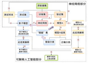
Figure 1. Workflow of the Shadow Learner System
| 神经网络部分 | Neural Network Module |
|
原数据集 |
Original dataset |
| 测试样本 | Test sample |
| 测试集 | Test set |
| 训练集 | Training set |
| 验证集 |
Validation set |
| 影子模型 |
Shadow model |
| 神经网络 | Neural network |
|
模型A |
Model A |
| 模型A之测试样本结果 |
Test sample results of Model A |
| 影子模型之测试样本结果 |
Test sample results of the shadow model |
| “错题”集 |
Error-case set |
| 模型A结果 |
Model A results |
| 此结果解释 |
Explanation of the results |
| 此结果解释 |
Explanation of the results |
| 错误分类 |
Misclassified |
| 正确分类 |
Correctly classified |
| 影子模型之解释可视化 |
Explanation visualisation by the shadow model |
| 模型A之解释可视化 |
Explanation visualisation by Model A |
|
可解释人工智能部分 |
Explainable AI Module |
| 解释可视化之复合图像 |
Composite image of explanation visualisation |
2. Design of the Dense_Inception Composite Neural Network
Based on DenseNet’s dense connection mechanism and InceptionV3’s advantages in multi-scale convolution, this project independently develops a Dense_Inception composite neural network. DenseNet strengthens gradient flow through feature reuse, while InceptionV3 uses asymmetric convolution to reduce computational cost while maintaining a large receptive field. The model is optimised for the characteristics of thyroid ultrasound images, such as low contrast, blurred boundaries and irregular nodule morphology, significantly improving feature extraction efficiency and classification accuracy. In the experimental design, the model will be compared with mainstream models such as VGG16, ResNet50, DenseNet and InceptionV3. Its accuracy, precision, recall and F1 score will be evaluated under both ImageNet pretraining and medical-image-only pretraining conditions, demonstrating the model’s superior performance in medical image processing tasks.
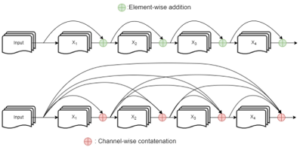
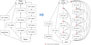
Figure 2. Schematic Diagram of the Dense_Inception Network Structure and Dense Connection Mechanism
3.Explainable AI Cross-Validation System Using Two Technologies
The system integrates two explainable AI technologies, Local Interpretable Model-agnostic Explanations (“LIME”) and XRAI, a region-attribution technology based on integrated gradients. These are used to visualise and cross-validate the decision results of both the standard model and the shadow model. LIME generates ROIs through super-pixel perturbation and offers advantages such as low computational cost and clear boundaries. XRAI combines integrated gradients with black-and-white baselines to provide stable and consistent attribution results. Using the two methods in a complementary manner can effectively avoid the problem of unstable interpretations associated with a single technology. Physicians can intuitively understand the basis of the model’s judgement and identify potentially error-prone areas through the highlighted ROIs in the images, thereby enhancing diagnostic trustworthiness and clinical acceptance. The system is designed in particular to assist with benign–malignant differentiation in patients with TI-RADS 3/4a thyroid nodules, helping some patients with a high likelihood of benign disease avoid unnecessary invasive needle aspiration biopsy. It therefore has significant clinical value and social relevance.
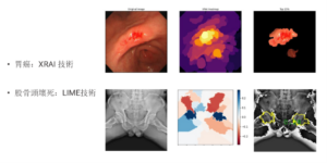
Figure 3. Example of Composite ROI Visualisation Generated by LIME and XRAI
| 胃癌:XRAI技術 |
Gastric Cancer: XRAI Technology |
|
股骨頭壤死:LIME技術 |
Femoral Head Necrosis: LIME Technology |
4. Error-Case Set Mechanism and Self-Iterative Model Optimisation
This project pioneers an “error-case set” mechanism, which systematically recycles ultrasound images misclassified by the standard model to form a dedicated shadow training set. The shadow model is then trained from scratch on this basis and remains completely independent of the original model weights, ensuring that its learned knowledge comes purely from error cases. Meanwhile, the error-case set can be fed back into the original standard model for advanced training, enabling self-iteration and continuous optimisation of the model and helping to break through the accuracy bottleneck of traditional deep learning models. This mechanism imitates the human cognitive process of “learning from mistakes” and provides a new path for closed-loop optimisation in medical imaging AI.
This project is dedicated to creating an intelligent medical imaging computer-aided diagnosis system with the ability to “learn from mistakes”. It overcomes the dual limitations of black-box operation and error discarding in existing CAD systems, and promotes the trustworthy application and implementation of AI in clinical diagnosis. By enhancing the accuracy and explainability of thyroid nodule diagnosis, the project supports the development of precision medicine and smart healthcare, creating new technical pathways and clinical models for future medical imaging AI and explainable AI.